Formulation and Evaluation of Nanoparticle-Based Drug Delivery Systems for Targeted Cancer Therapy
Keywords:
Nanoparticles, PLGA, targeted drug delivery, cancer therapy, nanoprecipitation, factorial design, drug release kinetics, entrapment efficiency, cytotoxicity, EPR effectAbstract
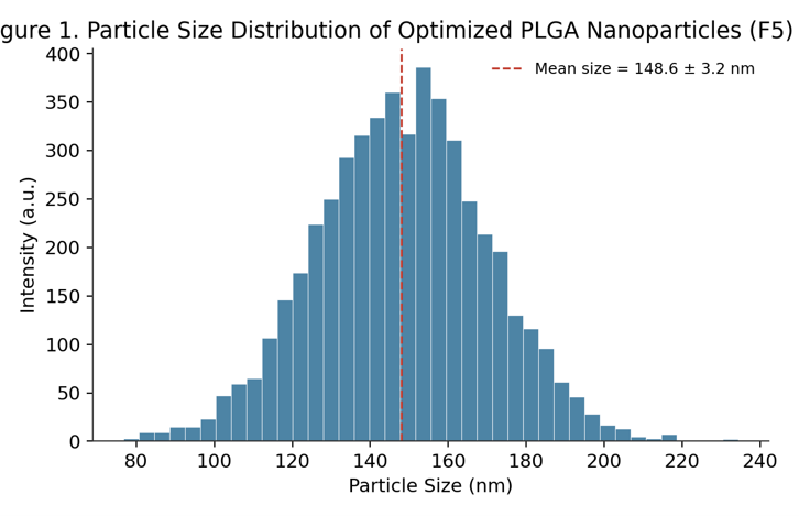
Traditional chemotherapy faces challenges such as the low water solubility of numerous anticancer drugs, non-specific distribution within the body, quick elimination from the system, and toxicity that limits the dosage due to harm to healthy tissues. Nanoparticle-based drug delivery systems (NDDS) have been developed as a promising approach to address these issues by allowing for controlled drug release, enhanced pharmacokinetics, and selective accumulation in tumors through passive (enhanced permeability and retention, EPR) and active (ligand-mediated) targeting methods. This study involved the formulation of poly(lactic-co-glycolic acid) (PLGA) nanoparticles containing a model anticancer drug (doxorubicin hydrochloride) using the nanoprecipitation (solvent displacement) method. The process was optimized with a 3² factorial design, considering polymer and surfactant concentrations as independent variables. Nine different formulations (F1–F9) were created and assessed for particle size, polydispersity index (PDI), zeta potential, entrapment efficiency (EE), drug loading (DL), surface morphology, in vitro drug release, release kinetics, and short-term stability. The optimized formulation (F5) showed a mean particle size of 148.6 ± 3.2 nm, a PDI of 0.181 ± 0.02, a zeta potential of −24.3 ± 1.1 mV, an entrapment efficiency of 78.4 ± 2.6%, and a drug loading of 8.9 ± 0.4%. Transmission electron microscopy revealed spherical, distinct particles with smooth surfaces. In vitro release experiments in phosphate-buffered saline (pH 7.4, 37 °C) exhibited a biphasic release pattern, with an initial burst (~12% within 2 hours) followed by a sustained release over 96 hours, which aligned best with the Korsmeyer–Peppas model (R² = 0.988, n = 0.41), suggesting Fickian diffusion along with polymer erosion. Cytotoxicity tests (MTT) on MCF-7 breast cancer cells showed that the nanoparticle formulation had significantly improved anticancer activity (IC₅₀ = 6.2 ± 0.5 µg/mL) compared to the free drug solution (IC₅₀ = 18.4 ± 1.2 µg/mL, p < 0.001), while blank nanoparticles exhibited minimal cytotoxicity, indicating the carrier's biocompatibility. Stability studies conducted under accelerated and refrigerated conditions over 90 days demonstrated that storage at 4 ± 2 °C maintained particle size, PDI, and entrapment efficiency much better than storage at 25 ± 2 °C/60% RH. These results suggest that PLGA nanoparticles are a viable, biocompatible platform for the targeted delivery of anticancer drugs, meriting further in vivo pharmacokinetic and tumor-targeting studies.
References
1. Danhier, F., Ansorena, E., Silva, J. M., Coco, R., Le Breton, A., & Préat, V. (2012). PLGA-based nanoparticles: An overview of biomedical applications. Journal of Controlled Release, 161(2), 505–522.
2. Barenholz, Y. (2012). Doxil® — The first FDA-approved nano-drug: Lessons learned. Journal of Controlled Release, 160(2), 117–134.
3. Matsumura, Y., & Maeda, H. (1986). A new concept for macromolecular therapeutics in cancer chemotherapy: Mechanism of tumoritropic accumulation of proteins and the antitumor agent smancs. Cancer Research, 46(12), 6387–6392.
4. Kumari, A., Yadav, S. K., & Yadav, S. C. (2010). Biodegradable polymeric nanoparticles based drug delivery systems. Colloids and Surfaces B: Biointerfaces, 75(1), 1–18.
5. Fonseca, C., Simões, S., & Gaspar, R. (2002). Paclitaxel-loaded PLGA nanoparticles: Preparation, physicochemical characterization and in vitro anti-tumoral activity. Journal of Controlled Release, 83(2), 273–286.
6. Mohanraj, V. J., & Chen, Y. (2006). Nanoparticles – A review. Tropical Journal of Pharmaceutical Research, 5(1), 561–573.
7. Ritger, P. L., & Peppas, N. A. (1987). A simple equation for description of solute release II: Fickian and anomalous release from swellable devices. Journal of Controlled Release, 5(1), 37–42.
8. Peer, D., Karp, J. M., Hong, S., Farokhzad, O. C., Margalit, R., & Langer, R. (2007). Nanocarriers as an emerging platform for cancer therapy. Nature Nanotechnology, 2(12), 751–760.
9. Bilati, U., Allémann, E., & Doelker, E. (2005). Development of a nanoprecipitation method intended for the entrapment of hydrophilic drugs into nanoparticles. European Journal of Pharmaceutical Sciences, 24(1), 67–75.
10. Zhao, Z., Ukidve, A., Kim, J., & Mitragotri, S. (2020). Targeting strategies for tissue-specific drug delivery. Cell, 181(1), 151–167.
11. Bazak, R., Houri, M., El Achy, S., Kamel, S., & Refaat, T. (2015). Cancer active targeting by nanoparticles: A comprehensive review of literature. Journal of Cancer Research and Clinical Oncology, 141(5), 769–784.
12. Makadia, H. K., & Siegel, S. J. (2011). Poly lactic-co-glycolic acid (PLGA) as biodegradable controlled drug delivery carrier. Polymers, 3(3), 1377–1397.
13. Anselmo, A. C., & Mitragotri, S. (2016). Nanoparticles in the clinic. Bioengineering & Translational Medicine, 1(1), 10–29.
14. Hans, M. L., & Lowman, A. M. (2002). Biodegradable nanoparticles for drug delivery and targeting. Current Opinion in Solid State and Materials Science, 6(4), 319–327.
15. Sinha, R., Kim, G. J., Nie, S., & Shin, D. M. (2006). Nanotechnology in cancer therapeutics: Bioconjugated nanoparticles for drug delivery. Molecular Cancer Therapeutics, 5(8), 1909–1917.
16. Panyam, J., & Labhasetwar, V. (2003). Biodegradable nanoparticles for drug and gene delivery to cells and tissue. Advanced Drug Delivery Reviews, 55(3), 329–347.
17. Costa, P., & Sousa Lobo, J. M. (2001). Modeling and comparison of dissolution profiles. European Journal of Pharmaceutical Sciences, 13(2), 123–133.
18. Chan, J. M., Valencia, P. M., Zhang, L., Langer, R., & Farokhzad, O. C. (2010). Polymeric nanoparticles for drug delivery. Methods in Molecular Biology, 624, 163–175.
19. Yoo, J., Park, C., Yi, G., Lee, D., & Koo, H. (2019). Active targeting strategies using biological ligands for nanoparticle drug delivery systems. Cancers, 11(5), 640.
20. Danhier, F. (2016). To exploit the tumor microenvironment: Since the EPR effect fails in the clinic, what is the future of nanomedicine? Journal of Controlled Release, 244, 108–121.
Additional Files
Published
Issue
Section
License
Copyright (c) 2026 International Journal of Fundamental Research & Development

This work is licensed under a Creative Commons Attribution 4.0 International License.